
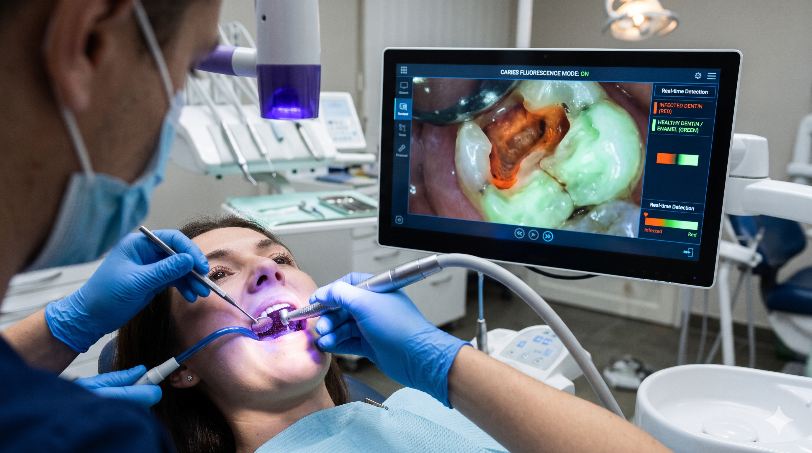
What If Dentists Could See Infected Dentin More Clearly While Removing Cavities?
How Could This Help Dentists?
- Identify Areas That Need Closer Examination
Fluorescence can highlight differences between dental tissues, providing dentists with additional visual information during cavity preparation.
- Support Conservative Caries Removal
By helping dentists assess the affected area, the technology may support more selective removal of carious tissue while preserving healthy tooth structure.
- Provide Visual Guidance During Treatment
Instead of relying only on colour, texture and tactile feedback, dentists can use fluorescence as an additional guide when assessing the cavity.
- Help Assess Remaining Carious Tissue
Fluorescence-based examination may help dentists identify areas that require further assessment before completing a restoration.
What Does This Mean for Dental Clinics?
Fluorescence-guided caries removal is an interesting development in minimally invasive dentistry.
For restorative dentists, it offers another way to assess carious tissue during treatment. However, fluorescence does not perfectly distinguish infected from healthy dentin, and the technique should complement established clinical criteria rather than replace them.